Laser Spectroscopy and
Nanoparticle Research
at The University of Texas in Austin
Nanoparticle Research
at The University of Texas in Austin

Bimaterial Heterostructures: Semiconductor core-shell nanoparticles studies
Bimaterial semiconductor Nanoparticles have recently
shown new physical phenomena such as enhanced Quantum Yield (QY) [1], which may be
useful in biophysics and labelling technologies (CdSe shelled ZnS
cores) [2].
Separation of Carriers, convenient for Lasing applications, has been
demonstrated as well, (ZnS shelled CdSe cores) [3].
 Fluorescence of the CdSe (dotted line) and CdSe-ZnS (solid line) nanocrystals[1] |
 Live Cell Imaging with Quantum Dots[2] |
 Light Amplification Using Inverted Core/Shell ZnSe/CdSe Nanocrystalls[3] |
In a new process for ZnS & CdSe core-shell nanoparticle production, a double ablation setup has been designed to produce a two step LAM process. In a first stage, spherical nanoparticles are generated. In a second cell they are mixed with microparticles of the second material, (that will form the shell), and the mixture
 is then ablated. The physics going on in this second ablation
of mixed micro and nanoparticles is particle size dependent.
Microparticles are
normally ablated as in the first
cell but nanoparticles (which have a diameter
comparable to the wavelength), on the other hand, are not expected to
develop a
shockwave. Because of the high temperature, (above the melting point
for ZnS
and CdSe), nanoparticles start to reduce they diameter, (in agreement
with
Thermodynamic calculations) and evaporated microparticles condense on
top of
them at the end of the laser pulse to form the shell.
is then ablated. The physics going on in this second ablation
of mixed micro and nanoparticles is particle size dependent.
Microparticles are
normally ablated as in the first
cell but nanoparticles (which have a diameter
comparable to the wavelength), on the other hand, are not expected to
develop a
shockwave. Because of the high temperature, (above the melting point
for ZnS
and CdSe), nanoparticles start to reduce they diameter, (in agreement
with
Thermodynamic calculations) and evaporated microparticles condense on
top of
them at the end of the laser pulse to form the shell. In the process of generating CdSe shells and ZnS cores, ZnS is ablated before mixing it with CdSe microparticles.
When generating ZnS shelled CdSe cores, we invert the order of the two ablation processes. CdSe is ablated before mixing it with ZnS microparticles.
Using a high resolution TEM, ZnS/CdSe LAM generated core/shell nanoparticles, were seen for the first time. In fact as far as we know these are the first core/shell particles of this kind got by any laser based production technique.
Based on TEM micrographs, the estimated mean core diameters range goes form 5 to 30nm. For the 20nm diameter cores, well formed shells were around 5nm tick. Furthermore, EDS measurements showed that core/shell structures were made of ZnS and CdSe respectively. FFT and Diffraction Patterns show cores and shells are crystals.
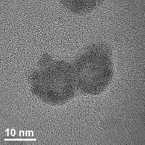
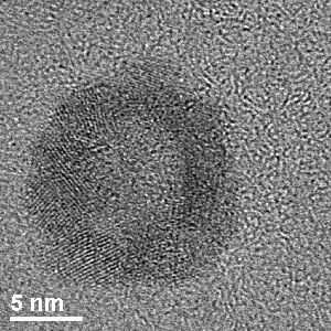
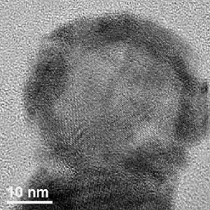
Under TEM conditions, the highest atomic number of CdSe makes the darkest images. ZnS is normally brighter than CdSe. This contrast is clearly seen in this pictures and it is supported by EDS measurements.
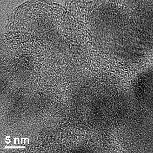
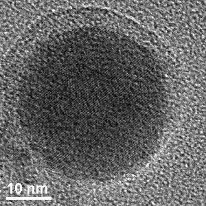
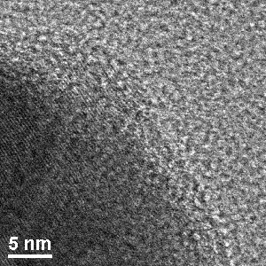
High resolution TEM micrographs of a CdSe-ZnS core-shell nanoparticle. The spherical core and covering shell can be observed. In the close-up the ZnS crystal planes can bee seen parallel to the core surface.
References:
[1] Synthesis and Characterization of Strongly Luminescing ZnS-Capped CdSe Nanocrystals; Guyot-Sionnest et al, J. Chem. Phys. 100, 468, (1996)
[2] Use of quantum dots for live cell imaging; Mattoussi et.al. Nature Methods 1, 73 (2004)
[3] Light Amplification Using Inverted Core/Shell Nanocrystals: Towards Lasing in the Single-Exciton Regime Klimov et al. J. Phys. Chem. B 108, 10625 (2004)